Handbook of EEG interpretation - part 9 ppt

Handbook of EEG interpretation - part 9 ppt
... Obstructive sleep apnea: implica-
tions for cardiac and vascular disease. JAMA 2003; 290 (14): 190 6– 191 4.
Shepard JWJ. ( 199 1). Atlas of Sleep Medicine. Futura, Mount Kisco, NY,
199 1.
Wittig RM, Zorick FJ, ... The
Report of an American Academy of Sleep Medicine Task Force. Sleep
199 ;22(5):667–6 89.
Arand D, Bonnet M, Hurwitz T, et al. The clinical use of the MSLT and MWT.
Sle...

Handbook of EEG interpretation - part 1 ppt
... bibliographical references and index.
ISBN-13: 97 8-1 -9 3 386 4-1 1-2 (pbk. : alk. paper)
ISBN-10: 1 -9 3386 4-1 1-7 (pbk. : alk. paper)
1. Electroencephalography—Handbooks, manuals, etc. I. Tatum, William ... absolute electrographic sites of maximal nega-
tivity (or positivity) by phase reversals (Figure 1.3A).
Normal EEG
7
Fp1-F7
F7-T3
T3-T5
T5-O1
Fp1-Ref
F7-Ref
T3-Ref
T5-...

Handbook of EEG interpretation - part 6 ppt
... partialis continua in a 41-year-old patient with sub-
jective tingling and “twitching” noted at the corner of the left side of the mouth.
Note the rhythmic delta frequencies on the EEG that phase reverse ... it is often use-
ful to change the paper speed from 10 mm/sec to 30 mm/sec for bet-
ter identification of sleep spindles.
CHAPTER 6
160
FIGURE 5.13. BiPLEDs in a 37-year-old HIV-...

Handbook of EEG interpretation - part 8 ppt
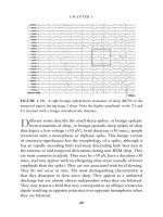
... figure above, bursts of 3- and 4-Hz dis-
charges (
arrows) were noted frequently during the PSG, particularly
in light stages of sleep.
Polysomnography
205
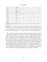
FIGURE 6.43. This is a 60-sec epoch demonstrating ... addi-
tion to the loss of alpha rhythm, there is appearance of slow,
rolling eye movements, mixed-frequency activity in the 2- to 7-Hz
range, and finally vertex waves.
Po...
Tài liệu Drugs and Poisons in Humans - A Handbook of Practical Analysis (Part 9) ppt
... acid-(t-BDMS)
2
15 69 2
1,4-dithiane 1068 11 69 1
thiodiglycol 1184 1468 1
thiodiglycol-TMS 1423 3
mustard sulfone 1433 1783 1
quinuclidin-3-ol (3-Q)-TMS 1267 2
benzilic acid-TMS 1 098 2
BZ-TMS 2633 2
N,N-diisopropylaminoethanol ... Remarks
DB-5* DB1701**
dimethyl methylphosphonate (DMMP) 881 (884) (1048) 1, (3)
O-ethyl-O-methyl methylphosphonate
(EMMP)
95 2 1112 1
O-isopropyl-O-methyl me...
Handbook of EEG interpretation - part 2 pdf
... theta rhythm in an 18-year-old patient
while awake.
T
heta rhythms are composed of 4- to 7-Hz frequencies of varying
amplitude and morphologies. Approximately one-third of nor-
mal awake, young ... voltages of >75 µV, while stage 4
consists of delta present for >50% of the recording.
Normal EEG
37
Activation techniques are a useful part of EEG in clinical practice...
Handbook of EEG interpretation - part 3 ppsx
... 1. 5- to 3.0-Hz delta in a 66-year-old man
with encephalopathy that was unresponsive. The above example of EEG is rep-
resentative of the entire record. No reactivity was noted during the EEG.
C
ontinuous ... the absence of IEDs in
the scalp EEG compared to the intracranial EEG where they occur at 1/sec.
I
t is often said that a normal interictal EEG does not exclude a cli...

Handbook of EEG interpretation - part 4 docx
... fragmentation of the
generalized discharge in the majority of cases.
CHAPTER 3
90
FIGURE 3.20. Slow-spike-and-wave in a patient with Lennox-Gastaut
syndrome.
S
low-spike(or sharp)-and-wave discharges ... figure.
CHAPTER 4
98
GENERALIZED SEIZURES
FIGURE 3.17. A burst of generalized 3.5-Hz spike-and-slow waves in JME.
S
pike-and-slow-wave complexes that have a repetition rate of &...

Handbook of EEG interpretation - part 5 potx
... 2001;18(5):442–455.
Verma A, Radtke R. EEG of partial seizures. J Clin Neurophysiol 2006;23:
333–3 39.
Westmoreland BF. The EEG findings in extratemporal seizures. Epilepsia
199 8; 39( Suppl 4):S1–S8.
CHAPTER ... drowsiness and light non-REM sleep. A frequent ictal pat-
tern of mesial temporal origin is the sudden appearance of localized
or regional background attenuation, build-u...

Handbook of EEG interpretation - part 7 potx
... arrow).
S
tage REM is characterized by the appearance of low-amplitude,
mixed-frequency EEG activity, EMG atonia, and rapid eye move-
ments. EEG activity is similar to that seen in stage I sleep; ... a 30-sec epoch showing the start of stage REM and
saw tooth waves.
S
aw tooth waves (arrows) are 2- to 5-Hz vertex negative sharp
waves that often occur in a series. They can be precu...
Từ khóa:
- the handbook of brain theory and neural networks
- handbook of couples therapy
- handbook of mechanical engineering
- reading upper intermediate part 9
- a history of britain part 9
- Báo cáo quy trình mua hàng CT CP Công Nghệ NPV
- Một số giải pháp nâng cao chất lượng streaming thích ứng video trên nền giao thức HTTP
- Nghiên cứu tổ chức chạy tàu hàng cố định theo thời gian trên đường sắt việt nam
- đề thi thử THPTQG 2019 toán THPT chuyên thái bình lần 2 có lời giải
- Giáo án Sinh học 11 bài 13: Thực hành phát hiện diệp lục và carôtenôit
- Giáo án Sinh học 11 bài 13: Thực hành phát hiện diệp lục và carôtenôit
- Giáo án Sinh học 11 bài 13: Thực hành phát hiện diệp lục và carôtenôit
- Giáo án Sinh học 11 bài 13: Thực hành phát hiện diệp lục và carôtenôit
- ĐỒ ÁN NGHIÊN CỨU CÔNG NGHỆ KẾT NỐI VÔ TUYẾN CỰ LY XA, CÔNG SUẤT THẤP LPWAN
- Phối hợp giữa phòng văn hóa và thông tin với phòng giáo dục và đào tạo trong việc tuyên truyền, giáo dục, vận động xây dựng nông thôn mới huyện thanh thủy, tỉnh phú thọ
- Phát hiện xâm nhập dựa trên thuật toán k means
- Nghiên cứu về mô hình thống kê học sâu và ứng dụng trong nhận dạng chữ viết tay hạn chế
- Nghiên cứu khả năng đo năng lượng điện bằng hệ thu thập dữ liệu 16 kênh DEWE 5000
- Sở hữu ruộng đất và kinh tế nông nghiệp châu ôn (lạng sơn) nửa đầu thế kỷ XIX
- Chuong 2 nhận dạng rui ro
- BT Tieng anh 6 UNIT 2
- Tranh tụng tại phiên tòa hình sự sơ thẩm theo pháp luật tố tụng hình sự Việt Nam từ thực tiễn xét xử của các Tòa án quân sự Quân khu (Luận văn thạc sĩ)
- chuong 1 tong quan quan tri rui ro
- Nguyên tắc phân hóa trách nhiệm hình sự đối với người dưới 18 tuổi phạm tội trong pháp luật hình sự Việt Nam (Luận văn thạc sĩ)
- Giáo án Sinh học 11 bài 14: Thực hành phát hiện hô hấp ở thực vật