Echocardiography A Practical Guide to Reporting - part 2 docx
Echocardiography A Practical Guide to Reporting - part 1 pot
... 4/5/07 12: 54 pm Page i
Ao aorta
ARVD arrhythmogenic right
ventricular dysplasia
ASD atrial septal defect
AV atrioventricular
AVSD atrioventricular septal
defect
BSA body surface area
ECG electrocardiogram
EOA ... minimum standard adult transthoracic study
Two-dimensional sections
• Parasternal long-axis.
• Parasternal long-axis views modified to show RV inflow and outflow.*
• Parasternal s...

Echocardiography A Practical Guide to Reporting - part 3 pps
... 91:1380 2.
6. Jenni R, Oechslin E, Schneider J, Attenhofer JC, Kaufmann PA. Echocardiographic and
pathoanatomical characteristics of isolated left ventricular non-compaction: a step
towards classification ... diagnosis of cardiomyopathy is made using all available clinical
data.
• The echocardiography report alone should never make a new diagno-
sis, but can suggest hypertrophic cardi...

Echocardiography A Practical Guide to Reporting - part 4 pdf
... how far down the aorta can flow reversal be detected on colour
mapping
Echocardiography: A Practical Guide for Reporting
42
ch05 4/5/07 1:33 pm Page 42
Echocardiography: A Practical Guide for Reporting
44
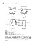
Figure ... commissures adapted from TOE. This can often be obtained transthoracically by
slight angulation and rotation from the apical 2- chamber view
ch05 4/5/07 1:33...

Echocardiography A Practical Guide to Reporting - part 5 ppsx
... Bileaflet
ch06 4/5/07 1:33 pm Page 65
Echocardiography: A Practical Guide for Reporting
58
Table 5.14 Echocardiography after mitral valve repair
• Appearance of the mitral valve and annuloplasty ... (b) Caged-ball mitral valve in a 4-chamber
view. (c) Tilting-disk aortic valve in a parasternal long-axis view. (d) Zoomed view of
a bileaflet mechanical mitral valve in a 4-c...

Echocardiography A Practical Guide to Reporting - part 6 pot
... chamber areas. Echocardiography 1984; 1:403 26 .
2. Davidson WR Jr, Pasquale MJ, Fanelli C. A Doppler echocardiographic examination
of the normal aortic valve and left ventricular outflow tract. ... al. ACC/AHA 20 06 guidelines for the management of patients with
valvular heart disease: a report of the American College of Cardiology/American Heart
Association Task Force on Practice Gu...

Echocardiography A Practical Guide to Reporting - part 8 potx
... diameter
throughout the respiratory cycle or after a sniff
ch 12 4/5/07 1:35 pm Page 111
Echocardiography: A Practical Guide for Reporting
116
3. LA or RA mass
• A mass attached to the atrial ... septum is likely to be a myxoma.
• A fixed mass attached away from the septum is likely to be malig-
nant. An associated pericardial effusion makes an angiosarcoma likely.
• F...

Echocardiography A Practical Guide to Reporting - part 9 pptx
... body surface
area (BSA)
a, 4
BSA (m
2
)
1.4–1.6 1.6–1.8 1.8 2. 0
1. Parasternal long-axis Diastole 3.4–4.9 3.6–5.1 3.9–5.3
Systole 2. 3–3.9 2. 4–4.1 2. 5–4.4
2. Parasternal short-axis, Diastole 3.7–5.4 ... pm Page 125
Echocardiography: A Practical Guide for Reporting
1 32
Figure A1 .3 Aortic dimensions by body surface area (BSA). (a, b) 95% range at the
sinus of valsalva for...

Echocardiography A Practical Guide to Reporting - part 10 pptx
... transoesophageal
echocardiography
transient ischaemic attack (TIA) 122
transmitral duration 13
transoesophageal echocardiography
(TOE)
adult congenital disease 99, 101
aortic dissection 82, 82
endocarditis ... orifice area
Appendices 4/5/07 12: 55 pm Page 135
Echocardiography: A Practical Guide for Reporting
136
Table A2 .2 Aortic position: Mechanical
V
max
Peak ∆P Mean ∆P EO...

Echocardiography A Practical Guide to Reporting - part 7 doc
... diagnosis (Tables 9 .2 and 9.3).
A guide threshold for RA dilatation is a transverse diameter >5 cm in
the 4-chamber view.
Table 9.1 LA dilatation
1 ,2
Mild
a
Moderate Severe
LA area (cm
2
) 20 29 30–40 ... chamber quantifica-
tion. Eur J Echocardiogr 20 06; 7:79–108.
2. Abhayaratna WP, Seward JB, Appleton CP, et al. Left atrial size: physiologic determi-
nants and clinical...
Từ khóa:
- a practical guide to linux commands
- intellectual property and open source a practical guide to protecting code
- essential guide to writing part 2
- a practical guide to solaris security
- a practical guide to protecting code
- a practical guide to fedora and redhat enterprise linux fifth edition odd answers
- chuyên đề điện xoay chiều theo dạng
- Nghiên cứu sự hình thành lớp bảo vệ và khả năng chống ăn mòn của thép bền thời tiết trong điều kiện khí hậu nhiệt đới việt nam
- Một số giải pháp nâng cao chất lượng streaming thích ứng video trên nền giao thức HTTP
- Nghiên cứu vật liệu biến hóa (metamaterials) hấp thụ sóng điện tử ở vùng tần số THz
- Biện pháp quản lý hoạt động dạy hát xoan trong trường trung học cơ sở huyện lâm thao, phú thọ
- ĐỒ ÁN NGHIÊN CỨU CÔNG NGHỆ KẾT NỐI VÔ TUYẾN CỰ LY XA, CÔNG SUẤT THẤP LPWAN
- NGHIÊN CỨU CÔNG NGHỆ KẾT NỐI VÔ TUYẾN CỰ LY XA, CÔNG SUẤT THẤP LPWAN SLIDE
- Phối hợp giữa phòng văn hóa và thông tin với phòng giáo dục và đào tạo trong việc tuyên truyền, giáo dục, vận động xây dựng nông thôn mới huyện thanh thủy, tỉnh phú thọ
- Nghiên cứu, xây dựng phần mềm smartscan và ứng dụng trong bảo vệ mạng máy tính chuyên dùng
- Tìm hiểu công cụ đánh giá hệ thống đảm bảo an toàn hệ thống thông tin
- Thơ nôm tứ tuyệt trào phúng hồ xuân hương
- Kiểm sát việc giải quyết tố giác, tin báo về tội phạm và kiến nghị khởi tố theo pháp luật tố tụng hình sự Việt Nam từ thực tiễn tỉnh Bình Định (Luận văn thạc sĩ)
- Quản lý nợ xấu tại Agribank chi nhánh huyện Phù Yên, tỉnh Sơn La (Luận văn thạc sĩ)
- BT Tieng anh 6 UNIT 2
- Tăng trưởng tín dụng hộ sản xuất nông nghiệp tại Ngân hàng Nông nghiệp và Phát triển nông thôn Việt Nam chi nhánh tỉnh Bắc Giang (Luận văn thạc sĩ)
- Tranh tụng tại phiên tòa hình sự sơ thẩm theo pháp luật tố tụng hình sự Việt Nam từ thực tiễn xét xử của các Tòa án quân sự Quân khu (Luận văn thạc sĩ)
- Giáo án Sinh học 11 bài 15: Tiêu hóa ở động vật
- Giáo án Sinh học 11 bài 15: Tiêu hóa ở động vật
- Giáo án Sinh học 11 bài 14: Thực hành phát hiện hô hấp ở thực vật
- Giáo án Sinh học 11 bài 14: Thực hành phát hiện hô hấp ở thực vật