Manual of Diagnostic Ultrasound in Infectious Tropical Diseases - part 4 pps

Manual of Diagnostic Ultrasound in Infectious Tropical Diseases - part 4 pps
... caval vein and a part of the
gall-bladder (b)
46 2 Typical Sonographic Findings in Inflammatory Diseases
Pyogenic abscesses caused by infectious organism via the bile ducts,
the portal vein, the ... normal find-
ing
56 2 Typical Sonographic Findings in Inflammatory Diseases
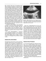
Fig. 2.55. In ammatory tumor. Con-
glomerate of in amed mesentery, in-
volved sections of the bowel,...

Manual of Diagnostic Ultrasound in Infectious Tropical Diseases - part 1 pps
... LiverTrematodeInfection(LiverDistosomiasis) 123
3.3.6 Schistosomiasis 130
3.3.7 Echinococcosis 143
4 Ultrasound F eatures in Childhood Infection 159
4. 1 UltrasoundinOsteomyelitis 159
4. 2 UltrasoundinBrainInfectioninNeonatesandInfants ... 165
4. 2.7 ViralEncephalitis 166
4. 3 UltrasoundinAbdominalandGastrointestinalInfections 166
Glossary 169
Suggested Reading 173
World Federation...

Manual of Diagnostic Ultrasound in Infectious Tropical Diseases - part 2 pptx
... early in
the exam
– check equipment settings again if findings are questionable
– repeat the examination within a short time in clinically difficult situa-
tions
1 .4
Interv en tional Ultrasound
In ... disadvantage of
dissolving rubber or plastic parts of the transducer.
22 2 Typical Sonographic Findings in Inflammatory Diseases
– emigration of leukocytes, neutrophils, or mono...

Manual of Diagnostic Ultrasound in Infectious Tropical Diseases - part 3 doc
... anterior mediastinum is scanned on both sides of the sternum.
34 2 Typical Sonographic Findings in Inflammatory Diseases
Table 2.2. Major infectious (tropical) diseases affecting the spleen
Tub ... pleura.
2.3 Organ-related Ultrasonic Findings 29
2.3.1 .4
Pathologic Findings
Lymph nodes are nearly always involved in inflammatory diseases, either
directly by the in fectious org...

Manual of Diagnostic Ultrasound in Infectious Tropical Diseases - part 5 pptx
... pathol-
ogy, but may indicate the presence of a non-neoplastic gastrointestinal
involvement.
In infectious diarrhea, ultrasound shows five sonographic patterns in
80% of cases:
1. Dilated intestinal ... sonographic signs of the intestinal walls of the small intestine and
colon: a typical finding in OI.
Fig. 3.19. Oblique scan passing through the left inferior quadrant of the...

Manual of Diagnostic Ultrasound in Infectious Tropical Diseases - part 6 ppt
... majority of patients overcoming either the classic or the hemor-
rhagic form of the disease remain in a considerably weak state for a period
of several weeks.
3.2.3 .4
Laboratory Findings
Findings include ... which is common in the tropical
and subtropical areas throughout the world, having its maximumincidence
at the end of the rainy season. A significant increase in the inci...

Manual of Diagnostic Ultrasound in Infectious Tropical Diseases - part 8 docx
... peripheral intrahep-
atic ducts from accompanying portal veins.
122 3 Ultrasound Diagnosis of Special Infectious and Parasitic Diseases
Fig. 3.57. Brazilian25-year-old male. Ul-
trasound of scrotal ... 132 3 Ultrasound Diagnosis of Special Infectious and Parasitic Diseases
Fig. 3.61. Macrosc opic examination of
the liver, show ing intense fibrous peri-
portal thickening
cir...

Manual of Diagnostic Ultrasound in Infectious Tropical Diseases - part 9 pdf
... in
neonates and infants can be clinically silent.
Once clinical suspicion is aroused and the physical examination and
laboratory tests are in keeping with an infectious process, plain film ra-
diographs ... a
needle inside the cyst.
b scolicide injection of
hyperosmolar saline so-
lution. c after injection.
d after reaspiration
152 3 Ultrasound Diagnosis of Special Infectious and...

General ultrasound In the critically ill - part 4 ppsx
... Radiologie
29 :46 9 -4 80
74 Chapter 12 Upper Extremity
Central Veins
Fig.
12.12.
Diaphanous curls are freely floating in the
lumen of the internal jugular vein. Since a part of this
image
...
Other Information Available from Abdominal
Aorta Study
For maximal use of the full potential of noninva-
sive ultrasound, it may be of interest to investigate
the aortic c...
General ultrasound In the critically ill - part 5 ppsx
... catheter.
Rean Soins Intens Med Urg
6:532
8. Lichtenstein D (19 94) Relevance of ultrasound in
predicting the ease of central venous line insertions.
Eur
J
Emerg 7 :46
9. Lichtenstein D, Saifi ...
- Using fluid therapy.
- Lowering the feet using the balance pedal of
the bed.
- Manually compressing the common femoral
vein at the groin (after checking its patency...
Từ khóa:
- infectious tropical diseases
- manual of diagnostic tests for aquatic animals 2009
- manual of diagnostic tests for aquatic animals 2013
- manual of diagnostic tests for aquatic animals
- manual of diagnostic tests and vaccines for terrestrial animals
- manual of diagnostic tests and vaccines for terrestrial animals 6th edition 2008
- Nghiên cứu tổ chức pha chế, đánh giá chất lượng thuốc tiêm truyền trong điều kiện dã ngoại
- Một số giải pháp nâng cao chất lượng streaming thích ứng video trên nền giao thức HTTP
- Nghiên cứu tổ chức chạy tàu hàng cố định theo thời gian trên đường sắt việt nam
- Giáo án Sinh học 11 bài 13: Thực hành phát hiện diệp lục và carôtenôit
- Giáo án Sinh học 11 bài 13: Thực hành phát hiện diệp lục và carôtenôit
- ĐỒ ÁN NGHIÊN CỨU CÔNG NGHỆ KẾT NỐI VÔ TUYẾN CỰ LY XA, CÔNG SUẤT THẤP LPWAN
- ĐỒ ÁN NGHIÊN CỨU CÔNG NGHỆ KẾT NỐI VÔ TUYẾN CỰ LY XA, CÔNG SUẤT THẤP LPWAN
- NGHIÊN CỨU CÔNG NGHỆ KẾT NỐI VÔ TUYẾN CỰ LY XA, CÔNG SUẤT THẤP LPWAN SLIDE
- Phát triển mạng lưới kinh doanh nước sạch tại công ty TNHH một thành viên kinh doanh nước sạch quảng ninh
- Nghiên cứu, xây dựng phần mềm smartscan và ứng dụng trong bảo vệ mạng máy tính chuyên dùng
- Nghiên cứu về mô hình thống kê học sâu và ứng dụng trong nhận dạng chữ viết tay hạn chế
- Định tội danh từ thực tiễn huyện Cần Giuộc, tỉnh Long An (Luận văn thạc sĩ)
- Tìm hiểu công cụ đánh giá hệ thống đảm bảo an toàn hệ thống thông tin
- Sở hữu ruộng đất và kinh tế nông nghiệp châu ôn (lạng sơn) nửa đầu thế kỷ XIX
- Chuong 2 nhận dạng rui ro
- Quản lý nợ xấu tại Agribank chi nhánh huyện Phù Yên, tỉnh Sơn La (Luận văn thạc sĩ)
- Tăng trưởng tín dụng hộ sản xuất nông nghiệp tại Ngân hàng Nông nghiệp và Phát triển nông thôn Việt Nam chi nhánh tỉnh Bắc Giang (Luận văn thạc sĩ)
- Giáo án Sinh học 11 bài 15: Tiêu hóa ở động vật
- Nguyên tắc phân hóa trách nhiệm hình sự đối với người dưới 18 tuổi phạm tội trong pháp luật hình sự Việt Nam (Luận văn thạc sĩ)
- Giáo án Sinh học 11 bài 14: Thực hành phát hiện hô hấp ở thực vật