Handbook of EEG interpretation - part 8 ppt

Handbook of EEG interpretation - part 8 ppt
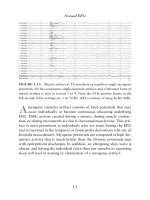
... figure above, bursts of 3- and 4-Hz dis-
charges (
arrows) were noted frequently during the PSG, particularly
in light stages of sleep.
Polysomnography
205
FIGURE 6.43. This is a 60-sec epoch demonstrating ... impedance mismatch between two elec-
trodes and compromises the common mode rejection ratio of the dif-
ferential amplifier. The result of this is presence of 60-Hz or othe...

Handbook of EEG interpretation - part 1 ppt
... bibliographical references and index.
ISBN-13: 97 8- 1 -9 3 386 4-1 1-2 (pbk. : alk. paper)
ISBN-10: 1-9 3 386 4-1 1-7 (pbk. : alk. paper)
1. Electroencephalography—Handbooks, manuals, etc. I. Tatum, William ... absolute electrographic sites of maximal nega-
tivity (or positivity) by phase reversals (Figure 1.3A).
Normal EEG
7
Fp1-F7
F7-T3
T3-T5
T5-O1
Fp1-Ref
F7-Ref
T3-Ref
T5...

Handbook of EEG interpretation - part 6 ppt
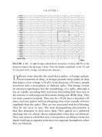
... 41-year-old patient with sub-
jective tingling and “twitching” noted at the corner of the left side of the mouth.
Note the rhythmic delta frequencies on the EEG that phase reverse at the F8
derivation.
T
he ... occur, and pro-
longed EEG recording can help elucidate the temporal pattern of patients with recur-
rent seizures when subtle or no clinical signs are present.
FIGURE 5. 18....

Handbook of EEG interpretation - part 9 ppt
... The
Report of an American Academy of Sleep Medicine Task Force. Sleep
199;22(5):667– 689 .
Arand D, Bonnet M, Hurwitz T, et al. The clinical use of the MSLT and MWT.
Sleep 22005 ;8( 1):123–144.
ASDA. EEG ... of the surgery, the patient is likely to have post-
operative hearing loss. However, the loss of the wave V is not incom-
patible with preserved hearing (false-positive). When...

Handbook of Economic Forecasting part 8 ppt
... squares (equation-by-equation, of course) estimator
of b, and the marginal density for is again of the inverted Wishart form. Symmetric
treatment of all equations is also feature of this formulation ... normal-Wishart prior, the ratio of the posterior variance of the “own”
lag coefficient in first equation to that of the “other” lag coefficient in second equa-
tion is identical...
Handbook of EEG interpretation - part 2 pdf
... theta rhythm in an 1 8- year-old patient
while awake.
T
heta rhythms are composed of 4- to 7-Hz frequencies of varying
amplitude and morphologies. Approximately one-third of nor-
mal awake, young ... predominance of non-REM appears in the first
part of the night, and REM in the last third of the night. A routine
EEG with REM may reflect sleep deprivation and not necessarily...
Handbook of EEG interpretation - part 3 ppsx
... 1. 5- to 3.0-Hz delta in a 66-year-old man
with encephalopathy that was unresponsive. The above example of EEG is rep-
resentative of the entire record. No reactivity was noted during the EEG.
C
ontinuous ... a night of ade-
quate sleep before demonstrating at least two naps with sleep-onset
REM is found in the context of the clinical history of excessive day-
time sleepiness...

Handbook of EEG interpretation - part 4 docx
... (petit mal) seizure in an 8- year-old boy.
D
uring an absence seizure, the EEG demonstrates generalized, reg-
ular, synchronous 3-Hz spike-and-wave discharges in the setting
of waking or drowsy background ... figure.
CHAPTER 4
98
GENERALIZED SEIZURES
FIGURE 3.17. A burst of generalized 3.5-Hz spike-and-slow waves in JME.
S
pike-and-slow-wave complexes that have a repetition rate of...

Handbook of EEG interpretation - part 5 potx
... drowsiness and light non-REM sleep. A frequent ictal pat-
tern of mesial temporal origin is the sudden appearance of localized
or regional background attenuation, build-up of 4- to 7-Hz rhythmic
activity, ... 2001; 18( 5):442–455.
Verma A, Radtke R. EEG of partial seizures. J Clin Neurophysiol 2006;23:
333–339.
Westmoreland BF. The EEG findings in extratemporal seizures. Epilep...

Handbook of EEG interpretation - part 7 potx
... arrow).
S
tage REM is characterized by the appearance of low-amplitude,
mixed-frequency EEG activity, EMG atonia, and rapid eye move-
ments. EEG activity is similar to that seen in stage I sleep; ... It must be remem-
bered that an increase in EMG without an EEG change cannot be
scored as an arousal, regardless the stage of sleep.
Polysomnography
185
FIGURE 6. 18. This is a 5-mi...
Từ khóa:
- handbook of shaft alignment part 13 pdf
- principles of marketing by philip kotler ppt chapter 8
- lego lord of the rings game walkthrough part 8
- tài liệu 1600 câu trắc nghiệm ôn thi hsg 8 part 14 pptx
- 8 overview of statistical interpretation of gwas
- 2012 guidelines for treatment of atopic eczema atopic dermatitis part i journal of the european academy of dermatology venereology 26 8 1045 1060
- the handbook of brain theory and neural networks
- handbook of couples therapy
- handbook of mechanical engineering
- handbook of english grammar
- the standard handbook of engineering calculations
- handbook of image and video processing
- the oxford handbook of ethical theory
- a handbook of commercial correspondence
- handbook of practical analysis
- Báo cáo thực tập tại nhà thuốc tại Thành phố Hồ Chí Minh năm 2018
- Báo cáo quy trình mua hàng CT CP Công Nghệ NPV
- chuyên đề điện xoay chiều theo dạng
- Nghiên cứu vật liệu biến hóa (metamaterials) hấp thụ sóng điện tử ở vùng tần số THz
- Biện pháp quản lý hoạt động dạy hát xoan trong trường trung học cơ sở huyện lâm thao, phú thọ
- Giáo án Sinh học 11 bài 13: Thực hành phát hiện diệp lục và carôtenôit
- Quản lý hoạt động học tập của học sinh theo hướng phát triển kỹ năng học tập hợp tác tại các trường phổ thông dân tộc bán trú huyện ba chẽ, tỉnh quảng ninh
- Phát triển du lịch bền vững trên cơ sở bảo vệ môi trường tự nhiên vịnh hạ long
- Phát hiện xâm nhập dựa trên thuật toán k means
- Nghiên cứu về mô hình thống kê học sâu và ứng dụng trong nhận dạng chữ viết tay hạn chế
- Tìm hiểu công cụ đánh giá hệ thống đảm bảo an toàn hệ thống thông tin
- Thiết kế và chế tạo mô hình biến tần (inverter) cho máy điều hòa không khí
- Sở hữu ruộng đất và kinh tế nông nghiệp châu ôn (lạng sơn) nửa đầu thế kỷ XIX
- Kiểm sát việc giải quyết tố giác, tin báo về tội phạm và kiến nghị khởi tố theo pháp luật tố tụng hình sự Việt Nam từ thực tiễn tỉnh Bình Định (Luận văn thạc sĩ)
- Quản lý nợ xấu tại Agribank chi nhánh huyện Phù Yên, tỉnh Sơn La (Luận văn thạc sĩ)
- BT Tieng anh 6 UNIT 2
- Tăng trưởng tín dụng hộ sản xuất nông nghiệp tại Ngân hàng Nông nghiệp và Phát triển nông thôn Việt Nam chi nhánh tỉnh Bắc Giang (Luận văn thạc sĩ)
- Giáo án Sinh học 11 bài 15: Tiêu hóa ở động vật
- Giáo án Sinh học 11 bài 14: Thực hành phát hiện hô hấp ở thực vật
- Giáo án Sinh học 11 bài 14: Thực hành phát hiện hô hấp ở thực vật